Resolving Renal Calculus Pain: Diagnostics, Solutions, and Choosing a Hospital

September 14, 2026 · 20 reads
Introduction
Few medical events disrupt daily life as abruptly as a kidney stone episode. Often starting as an ambiguous ache in the back or side, it can rapidly intensify into severe pain that leaves patients scrambling for immediate answers. Whether you are currently navigating an acute episode in an emergency setting or managing a history of recurrent stones, understanding how clinical decisions are made is crucial for taking control of your health.While renal stones are extremely common, treatment plans are never one-size-fits-all. Small mineral deposits often clear naturally with proper fluid management, but complex or obstructive stones require targeted surgical or non-invasive interventions. Navigating these choices during an uncomfortable episode can feel overwhelming. UrologyHospitals.com offers clear, patient-focused medical education to help you understand your symptoms, evaluate available clinical strategies, and connect with qualified specialists.
Understanding Renal Calculi: Physiology and Impact
Kidney stones, clinically referred to as renal calculi or nephrolithiasis, develop when dissolved minerals in your urine form solid crystalline structures within the renal system. Under normal conditions, natural compounds in urine stop these minerals from binding together. However, when urine volume drops or mineral concentration spikes, crystals settle, accumulate, and gradually form hard stones.
The urinary tract comprises four main linked structures: the kidneys, ureters, bladder, and urethra. The kidneys continually filter metabolic waste out of the bloodstream to form urine. As long as stones stay tiny and remain inside the kidney chambers, they may go completely unnoticed. Problems begin when a stone shifts into the ureter—the narrow tube connecting the kidney to the bladder. If a stone lodges there, it blocks urine flow, causes pressure to back up into the kidney, and triggers severe pain signals.
Recognizing Early Indicators and Warning Signals
Understanding how stone symptoms present allows you to seek timely medical intervention before severe urinary complications or kidney injury develop.
- Severe Flank and Abdominal Pain: Sharp, agonizing discomfort that begins below the ribcage along the back or side, frequently radiating downward into the lower stomach and groin.
- Colicky Pain Patterns: Discomfort that builds to a intense peak and then temporarily subsides, mirroring the rhythmic muscular contractions of the ureter trying to push the object downward.
- Dysuria and Urgency: A painful, burning sensation during urination, coupled with a constant, pressing feeling of needing to empty the bladder immediately.
- Hematuria (Visible or Microscopic Blood): Urine that appears pink, deep red, or brownish due to minor tissue abrasions caused by the stone moving against sensitive mucosal linings.
- Turbid or Malodorous Urine: Cloudy, unusually foul-smelling urine that can indicate cellular debris or an underlying urinary tract infection.
- Reflex Nausea and Upset Stomach: Nausea accompanied by vomiting, caused by shared neurological pathways between the digestive tract and the kidneys.
- Fever, Chills, and Systemic Distress: Clear signs that an infection has developed behind a urinary blockage, which constitutes a true medical emergency requiring immediate evaluation.
Because these symptoms overlap with other abdominal conditions—such as acute appendicitis, diverticulitis, or pelvic infections—a professional diagnostic evaluation is essential.
Underlying Drivers and Risk Profiles
Renal stones form due to an imbalance between fluid intake and mineral output. Identifying your specific risk profile helps guide both immediate management and long-term prevention strategies:
- Chronic Dehydration: Inadequate daily liquid intake concentrates urine, drastically increasing the likelihood of mineral precipitation.
- Dietary Sodium and Excess Animal Protein: High-salt diets increase the amount of calcium excreted by the kidneys, while heavy meat consumption raises uric acid levels and lowers urine pH.
- Familial Predisposition: Genetic factors heavily influence how your metabolic system processes calcium, oxalate, uric acid, and citrate.
- Metabolic Disorders: Health conditions such as hyperparathyroidism, gout, renal tubular acidosis, and insulin resistance alter urinary chemistry.
- Gastrointestinal Changes: Inflammatory bowel disease (IBD), chronic diarrhea, or prior bariatric surgery alter fat and water absorption, leading to elevated oxalate levels in the urine.
- Excess Body Mass: Higher BMI alters renal physiology and urinary pH, making stone formation more likely.
- Long-Term Medication Use: Certain prescription drugs, including specific diuretics, calcium-based antacids, and anti-seizure medications, can increase stone risk.
Clinical Diagnostics: Identifying Stone Characteristics
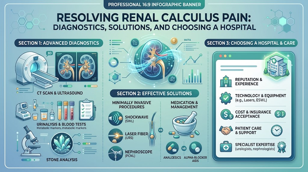
When you present with symptoms, healthcare providers run targeted diagnostic evaluations to establish the precise dimensions, composition, and location of the stone.
- Detailed Clinical History: Your provider documents symptom onset, pain patterns, family background, diet, and existing health conditions.
- Urinalysis and Culture: Laboratory evaluation detects microscopic blood, crystal types, pH levels, and bacteria indicating an active infection.
- Non-Contrast Helical CT Scan: The preferred, gold-standard imaging method for acute flank pain. It reveals virtually all stone types, precise millimeter measurements, anatomical location, and any secondary kidney swelling.
- Renal and Bladder Ultrasound: A safe, radiation-free imaging tool that visualizes kidney enlargement (hydronephrosis) and larger stones. It is especially useful for pregnant patients and children.
- Abdominal Radiography (KUB X-ray): A simple visual assessment tool used to track the movement of radiopaque calcium-containing stones over time.
- Comprehensive Metabolic Panel: Blood testing evaluates kidney function (creatinine and BUN), electrolyte levels, serum calcium, and uric acid concentration.
Comprehensive Spectrum of Care
Modern urological management balances effective stone clearance with minimal disruption to surrounding tissue. Specialists select therapies based on clinical urgency, anatomical factors, and stone characteristics.
Active Surveillance and Medical Expulsive Therapy (MET)
For smooth, small stones (less than 5mm) that do not obstruct urine flow:
- High-Volume Hydration: Consuming sufficient liquids to maintain clean, dilute urine output, helping naturally flush the stone out.
- Targeted Analgesia: Utilizing nonsteroidal anti-inflammatory drugs (NSAIDs) or alternative pain relievers to manage discomfort during the passing phase.
- Alpha-Blocker Medications: Prescription therapies that relax smooth muscle tissue in the lower ureter, allowing stones to move through faster with less pain.
Extracorporeal Shock Wave Lithotripsy (ESWL)
An outpatient, non-invasive treatment that focuses external acoustic shockwaves directly on a kidney stone to break it into tiny sand-like particles, which are then passed in the urine over the following days and weeks.
Ureteroscopy and Laser Lithotripsy (URS)
A minimally invasive procedure where a physician passes a microscopic scope through natural body pathways (urethra and bladder) directly into the ureter or kidney. A delicate laser fiber shatters the stone, and tiny fragments are removed using a specialized retrieval basket.
Percutaneous Nephrolithotomy (PCNL)
A specialized surgical procedure for large (greater than 20mm), unusually dense, or complex “staghorn” stones. The specialist creates a small keyhole incision in the patient’s back to insert a nephroscope directly into the kidney, using specialized tools to fragment and extract the stone.
Long-Term Metabolic Prevention
Once a stone is passed or surgically extracted, laboratory composition analysis combined with a 24-hour urine metabolic evaluation helps doctors design custom preventive plans, including dietary adjustments and specific preventative medications.
Overlapping Urological Conditions
Kidney stone disease rarely occurs in total isolation. Comprehensive medical evaluation often considers related urinary and pelvic health factors:
- Urinary Tract Infection Treatment: Trapped urine behind a blocked stone creates an environment where bacteria thrive rapidly. Resolving these infections requires prompt, targeted antibiotic treatment alongside stone clearance.
- Enlarged Prostate Treatment: In older men, benign prostatic hyperplasia (BPH) blocks normal bladder emptying, leading to stagnant urine that promotes bladder and kidney stone development.
- Prostate Cancer Treatment & Bladder Cancer Treatment: Blood in the urine is a classic symptom of kidney stones, but it can also be an early sign of prostate or bladder malignancies that require formal specialist evaluation.
- Robotic Urology Surgery: For complex underlying issues—such as ureteropelvic junction (UPJ) obstructions that cause recurrent stones—robotic-assisted surgical reconstruction provides precise tissue repair with reduced recovery times.
Evaluating Facilities and Specialists
Finding the right care team ensures you receive safe, effective, and personalized treatment. Use these clinical benchmarks when evaluating prospective providers:
- Specialist Qualifications: Seek out board-certified Urology Specialists who have focused training and experience in endourology and advanced stone management.
- Technology Infrastructure: Verify that the hospital features up-to-date diagnostic tools (high-resolution non-contrast CT and ultrasound) alongside advanced treatment capabilities (holmium/thulium lasers, ESWL technology, and robotic platforms).
- Emergency Response Readiness: Choose accredited Urology Hospitals that maintain 24/7 emergency departments with on-call urological surgical teams ready to handle acute blockages.
- Comprehensive Care Teams: Quality facilities coordinate care between urology, nephrology, interventional radiology, and nutritional science to offer complete stone prevention plans.
- Clear Patient Guidance: Look for teams that offer transparent discussions regarding procedure selection, potential complications, and realistic recovery timelines.
Optimizing Local Medical Searches
When researching local care options (often starting with a “Urologist Near Me” search), apply these criteria to narrow down your list:
- Confirm the specialist holds active surgical privileges at reputable regional medical centers.
- Check if the practice reserves open slots for urgent cases, ensuring you aren’t forced to wait weeks with an active stone.
- Ask about the clinic’s experience with the specific procedure recommended for your stone size and location.
- Ensure the facility accepts your health insurance or offers clear upfront pricing for self-pay options.
- Select practices that provide structured long-term follow-up care rather than short-term procedural fixes alone.
Side-by-Side Treatment Overview
| Interventional Category | Primary Clinical Indication | Primary Advantages | Clinical Considerations |
| Observation & MET | Small, non-blocking stones (< 5mm) | Completely non-invasive; low cost; managed at home | Requires patience; pain control; risk of unexpected impaction |
| Shock Wave Lithotripsy (ESWL) | Uncomplicated kidney stones (5–10mm) | Non-invasive; fast outpatient recovery; no incisions | May require secondary sessions; passing broken fragments can cause temporary pain |
| Laser Ureteroscopy (URS) | Ureteral & medium kidney stones (up to 20mm) | High clearance rate; direct visual access; no external cuts | Typically requires general anesthesia; temporary ureteral stent placement is common |
| Percutaneous Nephrolithotomy (PCNL) | Massive (> 20mm) or complex staghorn stones | Excellent clearance of large stone masses in one setting | Involves a surgical incision, brief hospital stay, and longer recovery period |
Hypothetical Clinical Scenarios
Scenario A: Managing an Uncomplicated Small Ureteral Stone
Case: A 38-year-old active adult develops sharp left flank discomfort. Diagnostic scanning reveals a 4mm stone in the mid-ureter, with no signs of infection or severe kidney strain.
Management: The specialist recommends conservative outpatient management with aggressive oral hydration, an alpha-blocker to relax the ureter, and target analgesics. The patient strains their urine and successfully passes the stone naturally within six days, avoiding surgery.
Scenario B: Resolving an Impacted Ureteral Stone
Case: A 56-year-old individual experiences persistent flank pain for over two weeks. Scans show an 9mm stone stuck in the lower ureter, causing moderate kidney swelling (hydronephrosis).
Management: The patient undergoes an outpatient ureteroscopy with laser lithotripsy. Using a flexible scope, the urologist breaks the stone into tiny dust particles and retrieves larger fragments with a basket, instantly relieving the blockage.
Step-by-Step Decision Framework
Step 1: Identify pain patterns and seek prompt diagnostic imaging.
  │
Step 2: Confirm exact stone dimensions, location, and check for infection.
  │
Step 3: Review non-invasive vs. surgical options with a urologist.
  │
Step 4: Balance procedure risks, success rates, and recovery times.
  │
Step 5: Select an accredited hospital and skilled urology specialist.
  │
Step 6: Undergo treatment and complete recommended follow-up appointments.
  │
Step 7: Complete a metabolic evaluation and adjust diet to prevent recurrence.
Critical Mistakes to Avoid
- Delaying Care for Persistent Pain: Ignoring prolonged flank discomfort can lead to unrecognized urinary blockages, severe infections, and long-term kidney damage.
- Assuming All Stones Need Surgery: Many small stones pass naturally with proper fluid intake, medication, and medical supervision.
- Relying on Self-Diagnosis: Assuming flank pain is just a simple muscle strain can delay diagnosis of serious internal blockages or appendicitis.
- Stopping Medications Early: Discontinuing prescribed antibiotics or ureteral muscle relaxants before your doctor advises increases your risk of recurring infection or stone impaction.
- Skipping Prevention Evaluations: Failing to run chemical stone analysis or 24-hour urine tests leaves you vulnerable to forming new stones in the future.
- Selecting Providers Based On Ads Alone: Choosing a hospital or surgeon based purely on promotional claims rather than verified credentials, facility capabilities, and specialist experience.
Essential Takeaways
- Timely diagnostic evaluation prevents silent kidney damage and severe systemic infections.
- High-resolution non-contrast CT scans offer the most accurate measurements of stone size and location.
- Treatment pathways range from active observation with fluid therapy to advanced laser and shockwave procedures.
- Choosing an accredited facility with experienced specialists ensures access to safe, modern treatments.
- Long-term prevention requires analyzing passed stones and addressing underlying metabolic factors.
Frequently Asked Questions (FAQs)
What does a urologist treat?
Urologists are medical specialists trained to diagnose and treat disorders of the male and female urinary system—including the kidneys, ureters, bladder, and urethra—as well as conditions affecting the male reproductive system.
When should someone see a urologist?
You should see a urologist if you experience severe back or side pain, visible blood in your urine, painful or difficult urination, recurrent urinary tract infections, or if imaging reveals structural issues in your urinary tract.
How are kidney stones diagnosed?
Urologists diagnose stones using a combination of physical exams, urine testing, blood panels, and targeted imaging. A non-contrast CT scan is the most accurate diagnostic tool for revealing stone size, shape, and location.
What treatments are available for kidney stones?
Care options range from simple fluid therapy and pain management for small stones to non-invasive Shock Wave Lithotripsy (ESWL), outpatient Laser Ureteroscopy (URS), and Percutaneous Nephrolithotomy (PCNL) for larger or complex stones.
What are common treatments for an enlarged prostate?
Benign prostatic hyperplasia (BPH) is managed through lifestyle changes, daily prescription medications, minimally invasive therapies (such as water vapor therapy or prostatic urethral lifts), or surgical options like TURP to clear blocked urine flow.
How is prostate cancer treated?
Treatment plans depend on cancer stage, risk group, and patient health. Options range from active surveillance and radical surgery to targeted radiation, hormonal therapies, and advanced systemic treatments coordinated by an oncology team.
When should urinary symptoms be evaluated?
Any sudden changes in urinary habits—such as severe pain, burning, an inability to urinate, or visible blood in the urine—require immediate evaluation by a healthcare professional to prevent complications.
What is robotic urology surgery?
Robotic urology surgery is a minimally invasive technique where a surgeon uses computer-controlled robotic arms to perform complex operations through tiny incisions, offering exceptional precision, minimal bleeding, and faster recoveries.
How can patients find a suitable Urology Hospital?
Look for accredited medical centers that offer comprehensive diagnostic tools, advanced surgical equipment, 24/7 emergency care, and board-certified urologists experienced in treating your specific condition.
How can someone find a Urologist Near Me?
Search medical board registries, ask your primary care physician for trusted referrals, verify hospital affiliations and insurance coverage, and ensure the specialist offers timely appointments for acute conditions.
Conclusion
Navigating a kidney stone episode can be daunting, but understanding how treatments are selected allows you to actively participate in your health decisions. Whether your stone requires simple fluid therapy under medical supervision or advanced laser lithotripsy, modern urological care offers safe, highly effective pathways to lasting relief. Partnering with a skilled specialist and choosing a well-equipped hospital ensures your care plan is tailored to your unique anatomical and health needs.Information platforms like UrologyHospitals.com help patients research conditions, understand modern procedures, and locate experienced healthcare providers. If you or someone you care for is experiencing symptoms of a kidney stone, schedule a clinical evaluation with a qualified specialist to begin your path to recovery.
Leave a Reply